Referring Doctors And Dentists
General

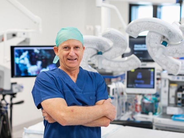
Dr Altmann is always happy to be contacted by doctors and dentists to give advice or to discuss current patients or patients yet to be referred.
Our preferred method of receiving referrals is by encrypted email.
We use Health Link and our edi is caltmann
Fax, mail or delivery by patient are other methods of referral delivery.
Generally blood investigation results are not required in the referral unless specifically relevant eg allergy testing and Dr Altmann prefers to view all relevant CT and MRI scan films on line with the patient present to explain their results.
We aim to send letters back to referring practitioners within 24 hours of patient’s consultation and encrypted email helps facilitate this.
Dr Altmann recognises the time pressures on doctors and dentists and strives to only include relevant information in his patient letters.
Please phone our receptionist Bev on (07) 4728 9886 if you have a patient you consider urgent and Bev can usually book that patient to be seen on the same day or next working day depending on the condition.
Urgent conditions include those where the patient is in acute pain due to otitis externa, otitis media, acute sinusitis, neck abscess or where you can see a head and neck malignancy or enlarged neck node.
Ear

Patients who require microscopic suction ear toilet for wax, debris or otitis externa can usually be booked for the same day or next working day if you phone Bev directly.
Otitis externa often results after water enters ears swimming or showering or after wearing earplugs or hearing aids. Staphylococcal infections are usually yellow while Pseudomonal infections are often bright green though this is by no means absolute.
Otitis externa often becomes fungal with candida albicans (white) and Aspergillus niger (black) often coexisting after previous treatment with antibiotics and broad spectrum ear drops.
Otocomb and Kenacomb ear drops are yellow, very thick and oily and often leave a thick residue on the tympanic membrane for months, causing reduced hearing and mimicking yellow pus.
Nose

Nasal Fracture
For a patient with a recent nasal fracture it is important for the referring doctor to examine the anterior nostrils to exclude a septal haematoma or abscess as these require early drainage. Although rare, septal haematomas are usually very obvious, appearing like a cherry involving the anterior nasal septum. It is also important to ask the patient about diplopia and check ocular movements in case there is a blow out fracture of the orbital rim associated with the nasal fracture.
For uncomplicated nasal fractures, no radiology is required. If complications such as coexistent orbital or zygomatic fracture, or septal haematoma are suspected then CT Sinuses/facial bones is all that is required, but should not delay referral.
Early referral of patients with nasal fractures is important. The best time for me to examine the patient’s nose is at around 1 week post injury, as swelling will have decreased, allowing deviation of the nasal bones to best be appreciated.
The best time to elevate the fractured nasal bones if they are deviated is at around 2 weeks post injury. Earlier than 2 weeks, the bones are often too mobile and won’t stay in place, and after 3 weeks the nasal bones have largely fixed and are much more difficult to manipulate though manipulation out to 4 weeks can still be attempted.
We perform all manipulations of fractured nasal bones under General Anaesthetic as this gives better patient comfort and allows a more precise result.
Rhinoplasty
Dr Altmann is happy to see all patients for rhinoplasty as long as there is a functional component to the condition. I don’t perform purely cosmetic rhinoplasty where there is no functional problem such as nasal obstruction or inability to wear safety glasses.
In order for rhinoplasty to attract a Medicare item number, there must be a history of trauma causing nasal deformity or congenital nasal deformity.
Throat

Acute
Acute presentations include embedded fish bones where there is always a history of eating fish with immediate pain and sensation of the fish bone catching. When a patient has a more gradual onset of throat discomfort and notes they have previously eaten fish, the diagnosis of embedded fish bone is very unlikely, although consideration needs to be given to an occult throat malignancy.
On examination, around 80% of embedded fish bones are in the tonsils or tongue base and are often visible under direct vision with good lighting.
The patient will often point externally to left or right upper neck which will help you find the bone. Please feel free to grasp the bone with Magill's or similar forceps and extract it in the direction it is pointing. The patient will always thank you immediately and tell you that their discomfort has gone once the bone is removed.
Quinsy is a condition which occurs occasionally and usually presents with acute, very lateralised ( left or right) throat pain and swelling, with varying degrees of trismus.
On examination the swelling is posterior and superior to the tonsil and usually there is minimal pus on the associated tonsillar surface. Quinsy can be difficult to discern from enlarged tonsils in EBV infection or bacterial tonsillitis.
More severe throat abscesses such as retropharyngeal abscesses need to be considered in unwell, febrile patients who are unable to swallow their own saliva. Always keep in mind the possibility of epiglottitis in adults and children who are unwell, febrile and drooling.
Chronic
Many malignancies of the upper aerodigestive tract will be visible in the oral cavity or oropharynx with good lighting.
Again the patient will often point externally and guide you to their tumour.
Enlarged neck lymph nodes can occur due to many causes such as congenital, infection and malignancy. Cat scratch disease and atypical TB should also be considered.
Lymph nodes in the neck should NOT be excised without full work up including full ENT examination, Fine Needle Aspiration cytology and CT neck.
We are happy to chat to referring doctors and dentists if they suspect their patient has an upper aerodigestive malignancy.
Get in Touch

Contact Details
Location
Suite 6, level 3, Mater Medical Centre
21-37 Fulham Rd, Pimlico
Townsville, QLD, Australia, 4812

Townsville, QLD, Australia, 4812